


"I was diagnosed with glaucoma during a routine eye exam. Dr. Johnson detected it early before I experienced any symptoms. With proper medication and regular follow-ups, my vision has remained stable for over 5 years now."
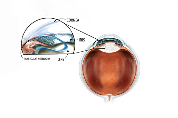
Glaucoma is a group of eye diseases that damage the optic nerve, often caused by increased eye pressure (IOP). If left untreated, it can lead to permanent vision loss. Glaucoma is one of the leading causes of blindness worldwide, but early detection and treatment can prevent further damage to the optic nerve and preserve vision.
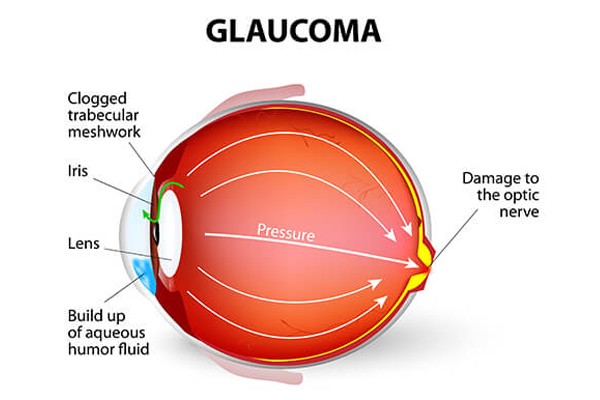
Glaucoma is often referred to as the "silent thief of sight" because it can develop without noticeable symptoms in its early stages. Regular eye exams are essential for early detection. The disease progresses slowly and can result in irreversible vision loss if not managed appropriately.
Early detection of glaucoma is critical for preventing vision loss. Watch this video to understand what happens as glaucoma progresses and why regular screening is important.
Once vision is lost to glaucoma, it cannot be recovered. Early treatment can preserve remaining vision and improve vision protection.
Glaucoma progresses slowly, making regular eye exams essential for early diagnosis.
Earlier treatment is typically more effective and may involve less invasive options.
Maintaining vision preserves independence and quality of life for patients.
While glaucoma may not present noticeable symptoms until significant damage has been done, there are some signs to be aware of, as well as risk factors that may increase your likelihood of developing the condition.
Having multiple risk factors significantly increases your risk. If you have several risk factors, consider more frequent eye examinations.
We use state-of-the-art technology to detect and monitor glaucoma, even in its earliest stages:
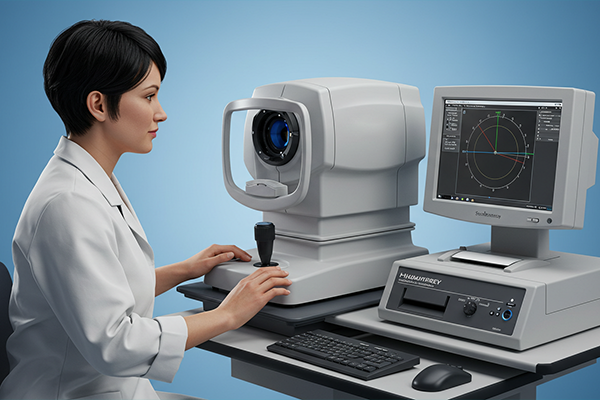
The Humphrey Visual Field Analyzer is the gold standard for detecting and monitoring vision loss due to glaucoma. This sophisticated test maps your entire field of vision, highlighting areas where vision may be compromised.
Benefits: Detects early vision loss before you notice symptoms, tracks progression over time, and helps evaluate treatment effectiveness.
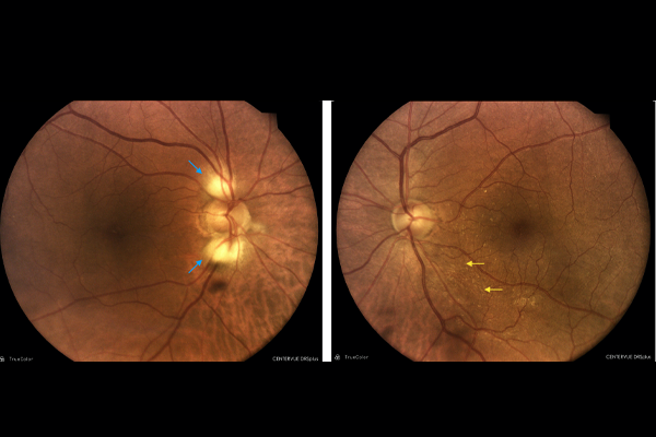
The RNFL scan uses Optical Coherence Tomography (OCT) technology to measure the thickness of the retinal nerve fiber layer. Thinning of this layer is often the earliest sign of glaucoma damage, occurring before visual field loss.
Benefits: Can detect glaucoma up to 5 years before visual field changes, provides objective measurements, and enables precise monitoring of disease progression.
When medications and laser treatments are not sufficient to control intraocular pressure, we offer several surgical interventions:
MIGS procedures are micro-scale interventions designed to lower eye pressure with fewer risks and a quicker recovery compared to traditional glaucoma surgery.
Ideal for: Patients with mild to moderate glaucoma, especially those also having cataract surgery.
Trabeculectomy is a traditional glaucoma filtration surgery that creates a new drainage pathway for excess fluid to leave the eye, effectively lowering intraocular pressure.
Ideal for: Patients with advanced glaucoma or those who continue to lose vision despite maximum medical therapy.
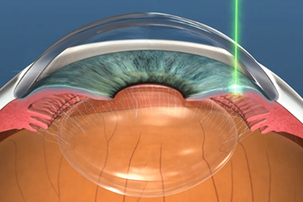
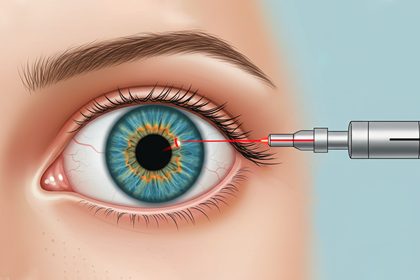
Laser Peripheral Iridotomy creates a small opening in the iris (the colored part of the eye) to improve fluid flow, particularly useful for angle-closure glaucoma or narrow-angle patients.
Ideal for: Patients with narrow angles or angle-closure glaucoma, often performed preventatively.
Glaucoma is a lifelong condition that requires ongoing care and monitoring. Our approach combines multiple strategies:
Prescription eye drops to lower intraocular pressure, often the first line of treatment. Different classes of medications work in various ways to reduce pressure.
Various laser procedures can improve drainage and lower pressure. Often used as an intermediate step between medications and incisional surgery.
Scheduled check-ups to monitor pressure, visual fields, and optic nerve appearance. Frequency depends on your condition's severity and stability.
Managing blood pressure, exercising appropriately, avoiding head-down positions for extended periods, and being careful with eye trauma risks.
Hear from patients who've benefited from our glaucoma care:
"I was diagnosed with glaucoma during a routine eye exam. Dr. Johnson detected it early before I experienced any symptoms. With proper medication and regular follow-ups, my vision has remained stable for over 5 years now."
"After struggling with multiple eye drops and side effects, I opted for MIGS surgery. The recovery was quick, and my pressure has been well-controlled since. I've even been able to reduce the number of medications I take daily."
"I experienced sudden eye pain and discovered I had angle-closure glaucoma. The team performed Laser PI immediately. Their quick response and excellent care prevented what could have been significant vision loss."
Schedule a comprehensive glaucoma screening today. Remember, most people with early-stage glaucoma have no symptoms.