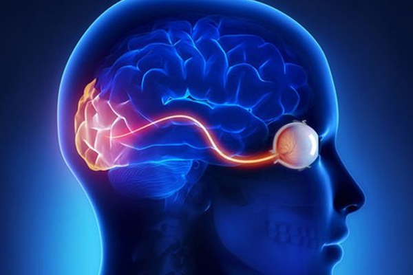
Neuro-ophthalmology is a specialized field that bridges neurology and ophthalmology, focusing on visual problems related to the nervous system rather than the eyes themselves.
Our neuro-ophthalmologists diagnose and treat visual issues caused by brain tumors, strokes, multiple sclerosis, and other neurological conditions that affect the complex visual pathways between the eye and brain. We specialize in optic nerve disorder and provide expert neuro eye care to patients suffering from vision impairments due to nervous system issues.
The human visual system is an intricate network that extends from the eyes through the optic nerves to the visual cortex in the brain. Damage at any point along this pathway can cause distinct visual problems that help locate the issue, including vision nerve disorders.
Our Neuro-ophthalmology specialists diagnose and treat a range of complex conditions where the visual and neurological systems intersect.
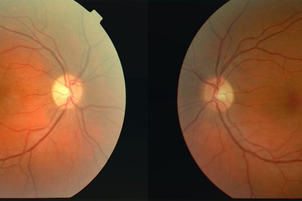
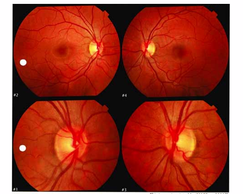
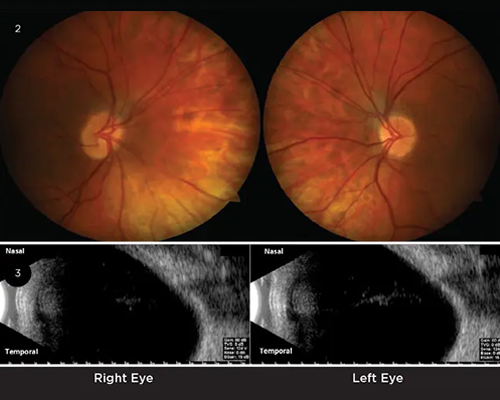
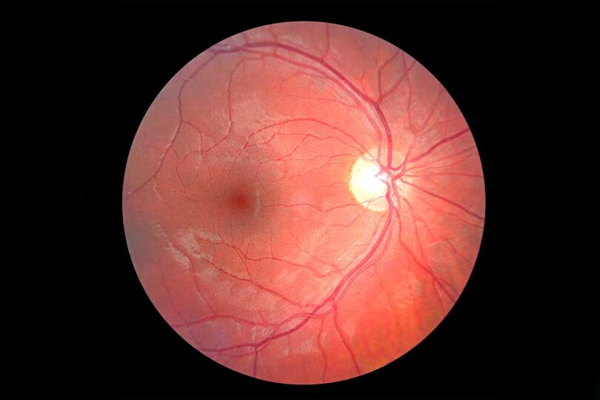
Optic neuritis is inflammation of the optic nerve, which carries visual information from the eye to the brain. This condition often causes sudden vision loss, eye pain (especially during eye movement), and may affect color perception.
Patient: 28-year-old female
Presentation: Sudden vision loss in right eye with pain during eye movement
Diagnosis: MRI revealed optic nerve enhancement consistent with optic neuritis, leading to MS diagnosis
Treatment: IV steroids followed by oral prednisone taper; referred to neurology for MS management
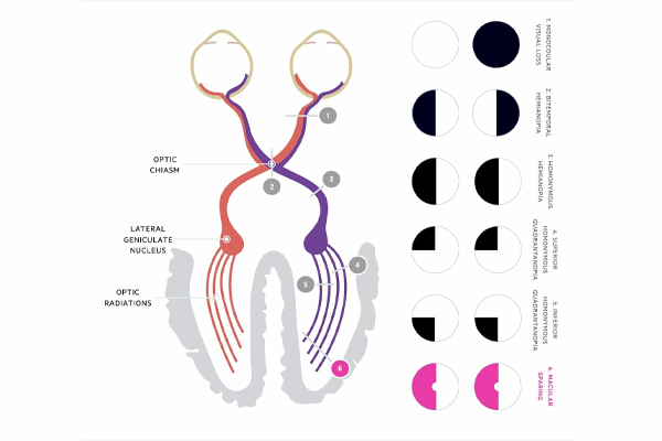
Visual field defects are areas of vision loss that can result from damage to the visual pathway at different points from the retina to the visual cortex in the brain. The pattern of vision loss often helps localize where the damage has occurred.
Patient: 65-year-old male
Presentation: Unable to see objects on his left side; had near-miss accidents while driving
Diagnosis: Right homonymous hemianopia due to left occipital lobe stroke
Treatment: Vision rehabilitation therapy and compensatory strategies; prism glasses to expand visual field
Cranial nerve palsies affecting vision involve damage to the nerves that control eye movement (oculomotor, trochlear, and abducens nerves - CN III, IV, and VI) and can cause double vision (diplopia), eye misalignment, and other visual disturbances.
Patient: 54-year-old with diabetes
Presentation: Sudden onset double vision, inability to look left
Diagnosis: Sixth nerve (abducens) palsy due to microvascular ischemia
Treatment: Better diabetes management; temporary prism glasses; condition resolved in 3 months
Neuro-ophthalmologic conditions require thorough evaluation using specialized tests to pinpoint the exact location and cause of the problem.
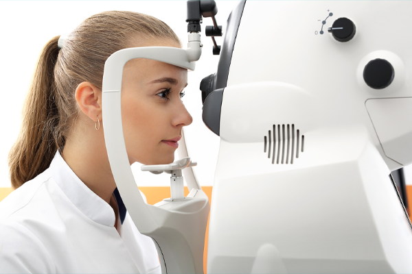
Measures peripheral vision and maps blind spots to help localize damage along the visual pathway.
Uses optical coherence tomography (OCT) to measure the thickness of nerve fiber layers in the retina and optic nerve.
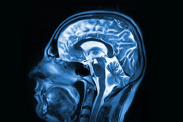
MRI scans with special protocols to visualize the brain, optic nerves, and visual pathways in detail.
Our multidisciplinary team tailors treatment to the specific condition and its underlying cause.
Our neuro-ophthalmologists develop individualized treatment strategies that address not only the visual symptoms but also the underlying neurological condition. This integrated approach ensures comprehensive care and the best possible visual outcomes.
Read what our patients have to say about their neuro-ophthalmology care and results.
"After my stroke, I couldn't see anything on my left side. The team not only diagnosed my condition precisely but gave me strategies and prism glasses that have helped me regain my independence."
"I woke up seeing double and was terrified. The doctor immediately recognized my cranial nerve palsy, explained how my diabetes had caused it, and reassured me it would likely be temporary. Three months later, my vision was back to normal."
"When I lost vision in my right eye, I was diagnosed with optic neuritis. This led to my MS diagnosis. The team coordinated with neurology and helped me understand how the conditions were connected and what to expect."
Our neuro-ophthalmology specialists are ready to help with complex visual disorders and provide answers to your vision concerns.


